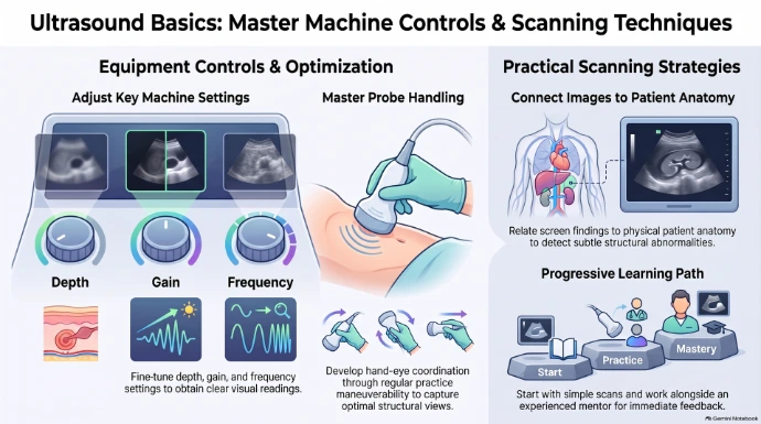
Stepping in front of an ultrasound machine for the first time can feel deeply intimidating. For medical trainees and beginners, confronting a complex console packed with knobs, buttons, and settings—while simultaneously attempting to interpret confusing gray-scale images on a screen—frequently leads to feeling overwhelmed and in over one's head. However, understanding the fundamentals of ultrasound is essential for delivering high-quality patient care.
Mastering clinical ultrasound is not an innate, secret talent reserved for a select few. Instead, it relies on a structured, deliberate approach that transforms equipment navigation and scan acquisition into second nature. With time, practice, and the right strategic mindset, any beginner can build clinical confidence behind the console.
1. Focus on the Big Three: Depth, Gain, and Frequency
For most beginners, equipment handling represents the very first major hurdle. Learning how to navigate the machine's controls while trying to optimize settings across varied ultrasound exam types can quickly induce cognitive overload. To build confidence and versatility across different clinical presentations, trainees must first master three fundamental wave parameters: depth, gain, and frequency.
"One of the biggest hurdles beginners face when learning ultrasound techniques is getting familiar with the equipment... understanding how to adjust the depth gain and frequency of the ultrasound waves is critical for obtaining clear images."
Different types of ultrasound exams demand tailored adjustments to achieve optimal image quality. Understanding how depth, gain, and frequency interact allows the clinician to efficiently adapt the machine settings to the specific anatomical target at hand. Mastering these core controls significantly reduces friction during a scan. When troubleshooting the equipment ceases to be a mental distraction, the operator can shift cognitive energy away from console frustration and focus entirely on acquiring clear, diagnostic views of organs and underlying structures.
2. Conquer Hand-Eye Coordination with Simple Scans and Feedback
Beyond manipulating the console, trainees face immediate physical challenges on the patient side of the machine. Maneuvering the probe effectively to capture optimal views requires fine hand-eye coordination that takes dedicated practice to develop. Furthermore, image interpretation requires experience to reliably distinguish normal findings from subtle abnormalities.
To systematically build tactile skill, optimize images, and master probe mechanics, beginners should adopt a two-part practical strategy:
- Progress from Simple to Complex Exams: Begin by mastering standard, straightforward imaging techniques before attempting more intricate or specialized exams. Building a foundation on routine scans prevents early frustration and establishes solid core mechanics.
- Practice Directly Alongside a Mentor: Perform scans in the direct presence of an experienced sonographer or mentor who can evaluate your physical technique and provide immediate, real-time feedback.
Relying on solo trial-and-error often leads beginners to establish bad motor habits and incorrect probe mechanics early in training—flaws that become deeply ingrained and far more difficult to unlearn later. Immediate feedback from an experienced mentor acts as a crucial preventative measure. It accelerates muscle memory, helps trainees rapidly develop an "eye" for image quality, and teaches them how to adjust wave parameters efficiently for any given exam.
While mastering equipment handling and fine motor control builds physical competence, reaching true efficiency requires transitioning from manual mechanics to cognitive perspective.
3. Stop Staring Only at the Screen—Focus on Patient Anatomy
Perhaps the single most transformative technique for a novice scanner is a fundamental shift in mindset: moving primary focus away from screen pixels and re-centering it directly on the physical patient anatomy.
"Focus on the patient's anatomy instead of just looking at the images on the screen... this technique takes practice but it's a gamechanger for beginners looking to take their ultrasound skills to the next level."
Instead of blindly hunting for abstract gray shapes on a monitor, take a moment to conceptualize the patient's actual anatomical structures and how they relate spatially to the transducer and the resulting screen display. This conceptual pivot moves a beginner from blindly searching for patterns to actively mapping real-world human anatomy in real time. By continually grounding the screen image in the patient's physical anatomy, scanners gain a far deeper understanding of the structures in view, enabling them to pick up on subtle details and identify abnormalities that might otherwise go completely unnoticed.
Conclusion
Conquering ultrasound basics requires a balanced approach centered on core equipment control, deliberate mentored practice, and an anatomy-first mindset. By mastering core settings like depth, gain, and frequency across different exam types, refining probe mechanics alongside an experienced mentor, and looking through the screen to focus on the physical patient, any beginner can cultivate complete confidence behind the machine.
As you approach your next clinical scan, reflect on your current workflow: Are you simply hunting for gray shapes on a monitor, or are you actively using your machine to map the underlying human anatomy?
Step Up Your Game
Achieve clinical excellence, master advanced imaging techniques, and build diagnostic confidence with our expert-led curriculum, designed to empower every sonographer’s journey toward mastery.